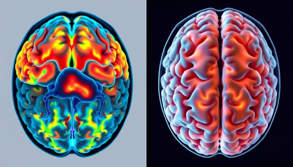
Did you know about 150 million people are facing Depressive Disorder around the world? This fact shows we need to understand mental health better, especially anxiety disorders. Brain scans from people with anxiety vs. those without tell an interesting story. They show how anxiety changes the way the brain works.
Recent fMRI studies found that people with social anxiety use a different part of the forebrain when dealing with threats or rewards. This means they think differently when making decisions. These studies give us a peek into how anxious people might have trouble controlling their emotions. They may also avoid being around others more often. With these findings, we can hope for better treatments for anxiety based on how it changes brain activity.
Key Takeaways
- Approximately 150 million people globally suffer from Depressive Disorder.
- Anxious individuals show differences in forebrain activation compared to non-anxious individuals.
- Brain imaging research reveals that anxiety can affect emotional control significantly.
- Different brain areas are activated during decision-making processes for those with anxiety.
- Understanding these neurological differences can lead to improved therapies for anxiety disorders.
Understanding Anxiety and Its Impact on the Brain
Every year, anxiety disorders affect millions of Americans. They reduce the quality of life. These conditions include types like generalized anxiety disorder and social anxiety. Knowing more about anxiety helps us find better treatments.
Defining Anxiety Disorders
Anxiety disorders mean you worry a lot and feel scared of things that might not be dangerous. Symptoms include being restless, getting irritated easily, and having trouble focusing. Things like hormone problems and stress can cause these disorders. Knowing the difference between normal worry and an anxiety disorder is important.
Common Symptoms of Anxiety
- Excessive worrying
- Restlessness or feeling on edge
- Irritability
- Fatigue
- Difficulty concentrating
- Sleep disturbances
It’s important to recognize these symptoms early. This can help understand how anxiety affects your brain. Experts in brain imaging can show us how anxiety changes brain responses and feelings.
The Importance of Brain Function in Anxiety
The brain plays a key role in anxiety. Problems in areas like the amygdala can make anxiety worse. Research on the brain shows patterns that can help with treatment. The J. Flowers Health Institute uses this information to make personalized wellness plans.
The impact of anxiety on the brain shows why early help and the right therapy matter. Knowing how brain function and anxiety symptoms are linked helps people find better treatment. This improves mental health.
Neuroimaging and Its Role in Anxiety Research
Neuroimaging tools help us understand anxiety disorders better. They let researchers see how the brain works when facing anxiety. One key method is functional Magnetic Resonance Imaging, or fMRI. It’s great for finding brain patterns related to anxiety in certain situations.
Introduction to Neuroimaging Techniques
Neuroimaging includes different ways to look at the brain’s structure and function. With CT and MRI scans, we learn about anxiety’s neuroanatomy. Studies show anxiety involves key areas like the limbic system and prefrontal cortex. This shows how complex anxiety and its brain connections are.
Functional MRI (fMRI) and Its Applications
fMRI has changed how we study anxiety with live brain activity views. It shows the amygdala’s increased activity during fear situations. The prefrontal cortex and hippocampus are also key in handling anxiety. fMRI helps explore how different brain areas relate to anxiety, leading to better treatments.
Understanding Brain Activation Patterns
Seeing how the brain activates is key in anxiety disorder research. Imaging shows special activity in places like the amygdala when anxious. Rest-state fMRI also links brain activity with anxiety traits. These findings help spot abnormal brain activity for better anxiety treatments.
What Happens in the Brain During Anxiety
Anxiety sets off complex reactions in the brain. It mainly involves areas key to processing emotions. By understanding these areas, we get insight into anxiety disorders. We see what changes in brain regions in anxiety mean for people with these disorders.
Brain Regions Most Affected by Anxiety
Some parts of the brain are more affected by anxiety. These include:
- Amygdala: Central to fear processing, it triggers the fight-or-flight response.
- Insula: It helps in emotional awareness and feeling bodily sensations.
- Anterieur Cingulate Cortex (ACC): It plays a role in managing emotions and feeling pain.
These areas together form the “fear network.” Their connection is key in how anxiety works in the brain.
Neural Correlates of Anxiety Disorders
Understanding these correlates helps in analyzing anxiety disorders. Research has found:
- Specific phobia affects 12.1% of people in a year, with the amygdala reacting strongly to threats.
- Social anxiety disorder has a 7.4% rate, linked to changes in the insula and ACC.
- Panic disorder is seen in about 2.7% of U.S. adults. Brain scans show the ACC is very active during panic attacks.
In both kids and adults, knowing the neural correlates is key for treatment. For more on this topic, read the full study on anxiety and the brain.
| Anxiety Disorder | 12-Month Prevalence Rate (%) | Key Brain Regions Involved |
|---|---|---|
| Specific Phobia | 12.1 | Amygdala |
| Social Anxiety Disorder | 7.4 | Insula, Anterior Cingulate Cortex |
| Panic Disorder | 2.7 | Anterieur Cingulate Cortex |
| Agoraphobia | 2.5 | Various Regions (Less Studied) |
Anxiety Brain Scan vs Normal: Evaluating Differences
Recent studies using brain imaging show big differences between anxious and healthy brains. Techniques like functional magnetic resonance imaging (fMRI) let researchers see how the brain works. This includes looking at anxiety disorders.
Key Findings from Recent Studies
A study looked at people with Generalized Anxiety Disorder (GAD) and healthy individuals. It compared 16 people with GAD to 17 without it. The research showed key differences in brain connections by using fMRI data. This data also included 31 more healthy people.
People with GAD had weaker connections in certain parts of the brain compared to those without GAD. Their brains didn’t link up as well in areas judging what’s important. Yet, they had stronger connections in parts managing emotions.
Comparative Analysis of Brain Scans
The comparison of normal vs anxious brain scans highlights the different structures and functions in anxious brains. EEG studies also showed unique patterns. For example, GAD patients had more gamma band power, linked to high anxiety. Other anxiety disorders showed different patterns, like lower motivation signals.

These brain imaging findings help us understand mental health better. They show what happens in the brain with anxiety. This knowledge guides new treatments. It also encourages more research into the complex world of anxiety and the brain.
Brain Activation Patterns in Anxiety Disorders
When we look at brain activation in those with anxiety disorders, we find something interesting. Studies highlight a key player: enhanced amygdala activation. This part of the brain deals with emotions and fear. It’s often too active in people with anxiety.
Increased Amygdala Activation
Many studies point out the amygdala’s extra activity, especially with generalized anxiety disorder (GAD). Such an increase links to being more sensitive to scary things, causing stronger fear reactions. Neuroimaging helps see that those with more GAD show this increased activity clearly when facing fearful situations.
Role of the Insula in Anxiety Responses
The insula also matters a lot for anxiety. It helps connect how we feel emotionally with how our body feels. People with anxiety have different insula activity when they see something that scares them. This likely makes them more aware of body feelings, adding to their anxiety. It helps us see how the brain’s networks work together in anxiety, making our understanding of it better.
Biological Markers of Anxiety: Understanding the Science
Biological markers of anxiety are key to understanding and diagnosing anxiety disorders. They are biochemical indicators found through tests like neuroimaging. Recognizing these markers helps find the causes of anxiety and create better treatments.
What are Biological Markers?
Biological markers show a body’s physiological state. For anxiety, this includes hormone levels and neurotransmitter concentrations. Studies show cortisol and neurotransmitter levels relate closely to anxiety symptoms. These markers help us see how anxiety works and affects people.
Identifying Markers Through Brain Imaging
Brain imaging provides insights into anxiety’s biological markers. It allows researchers to see the brain’s structure and how it functions. This way, they find markers that show unusual activity in anxious brains.
Studies show that cortisol and melatonin levels change during anxiety. Low cortisol is common in long-term anxiety. Brain imaging also helps understand neurotransmitter activity, like serotonin, in anxiety.

| Biological Marker | Indicator of Anxiety | Relation to Anxiety Disorders |
|---|---|---|
| Cortisol | Stress hormone levels | Lower levels often found in chronic anxiety cases |
| Melatonin | Sleep-related hormone | Reduced levels in individuals with PTSD |
| Serotonin | Neurotransmitter | Lower plasma levels in panic disorder patients |
| Salivary Alpha-Amylase | Digestive enzyme linked to stress | Increased with heightened anxiety |
Studying these markers helps researchers and doctors better understand and treat anxiety disorders. This leads to more accurate diagnoses and effective treatment plans.
Clinical Implications of Anxiety Brain Scanning
Anxiety disorders are tough to diagnose and treat. Using brain scanning could change the game for mental health experts. These scans offer views into the brain’s workings, improving how we assess and treat anxiety.
Using Brain Scans for Diagnosis
Brain scans give a clearer view of anxiety disorders. They show specific patterns in the brain. This helps tell different anxieties apart, like PTSD. Knowing these patterns can make diagnosis more precise and personalize care.
Potential for Personalized Treatment Options
Brain scans could make anxiety treatment more personal. Treatments can match a person’s brain pattern. This might make care more effective, easing symptoms better. Therapists could use this info to find the best treatments, from talk therapy to medicines.
The Neuroscience of Anxiety Disorders
Understanding the neuroscience of anxiety unveils how fear impacts our feelings. Scientists have pinpointed key brain areas linked with anxiety. They have shed light on fear reactions and the crucial role of the anterior cingulate cortex. These discoveries could lead to new treatments.
Understanding Fear Circuits in the Brain
The fear circuits in our brain are key to how we react to scary situations. When we feel anxious, the amygdala gets activated. This makes our emotions more intense.
This activation is tied to how we see threats. It can cause too much anxiety in some people. Also, the dorsal anterior cingulate cortex processes our emotional responses. It helps control our fear. When these circuits don’t work right, anxiety disorder symptoms can get worse.
Role of the Dorsal Anterior Cingulate Cortex
The dorsal anterior cingulate cortex merges our feelings and thoughts. It plays a part in evaluating risks and managing emotional reactions. This affects how we handle anxiety.
Research on people with generalized anxiety disorder (GAD) has shown changes in this brain area. Studies with over 1,000 GAD patients found differences in how this cortex works. This highlights its role in anxiety disorders. These insights are key to creating new therapies.

Research Findings: Neuroscience of Anxiety
Recent studies using advanced neuroimaging techniques have given us deep insights into anxiety. These studies show consistent brain activity patterns. This helps us understand emotional dysregulation in anxiety disorders better. By combining data from various studies, researchers have found important differences in how the brain works in those with anxiety.
Meta-Analysis of Brain Activity Studies
Brain activity meta-analysis has shown us how anxiety appears in the brain. Researchers Alexander Shackman and Juyoen Hur have looked into how fear and anxiety share brain circuits. They found that the same areas, like the amygdala and the BNST, are active when people expect threats. This challenges old ideas and shows the complexity of brain activity in anxiety. It suggests we should view anxiety disorders as a range of related issues.
Impact of Emotional Dysregulation in Anxiety Disorders
Emotional dysregulation in anxiety greatly affects how people handle stress. A study by the University of Trento found differences in the brains of people with different types of anxiety. People with chronic anxiety disorders have changes in their anterior cingulate cortex. This part of the brain is key in controlling emotions. These findings could lead to new ways to diagnose and treat anxiety.
Conclusion
Looking at brain scans, we can see clear differences between people with and without anxiety disorders. The comparison of an anxiety brain scan vs normal shows major changes in how the brain works. This is especially true for key areas like the amygdala and prefrontal cortex.
These findings highlight the need for personalized methods to diagnose and treat anxiety. Doing so aims to improve the health of those affected.
It’s crucial to keep studying anxiety to learn more. Future research will help us understand the brain processes behind these disorders. By including more people in studies, scientists can uncover the layers of complexity in anxiety.
One study funded by the National Institutes of Health showed interesting results. It found that anxious children have lower brain signal changes across certain brain areas. This points to a complex mix of underlying issues.
Continuous research into how the brain functions in anxiety can lead to new ways to find and treat it. As we learn more, combining techniques like fMRI and detailed brain analyses will offer new insights. This could greatly help people living with anxiety, making their future brighter.
If you’re keen to know more about the latest in mental health and brain scans, here’s a helpful article on brain research.